
Stahls, Tags & Ear Rarities
There are a number of ear conditions which are obvious at birth. Some require surgery if a correction is desired, and others can be corrected without surgery at birth if splints are applied early enough.
Cryptotia, Stahl's bar, conchal crus and anteverted concha are ear conditions obvious at birth which can be corrected without surgery using EarBuddies™ Splints, either by parents at home, or with the aid of a Professional Fitter. If the opportunity to use EarBuddies™ is missed, then surgery is an option.
Splintage is not appropriate for the correction of pre-auricular tags, for Darwin's tubercles, or for ear clefts, pits or sinuses, and if a correction is desired, then formal surgery is required.
Some pre-auricular tags can be treated with a clipping method which does not require general anaesthesia, in this treatment is undertaken in the first few weeks after birth.
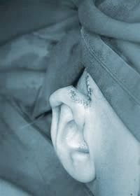
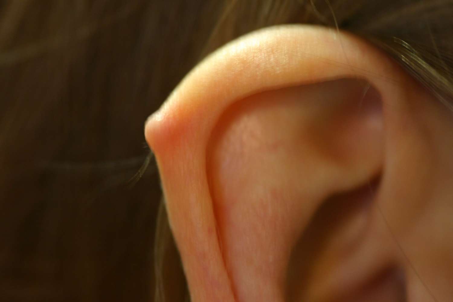
STAHL'S BAR
Also known as a Stahl's Ear, third crus or Spock Ear, A Stahl’s Bar is a frequent finding at birth, and can affect one or both ears. The bar can often give the ear a pointed appeance, sometimes called an Elf or Elfin ear.
If is possible to correct the condition early by splinting with EarBuddies™ Splints; if the opportunity is missed, then surgery is possible, preferably after the age of at least five years.
Surgical correction can be difficult. A direct wedge excision of the Stahl’s bar [skin and cartilage] is possible but can leave a visible scar in some skins particularly. A reconstruction using cartilage from the chest often gives excellent and reliable results.
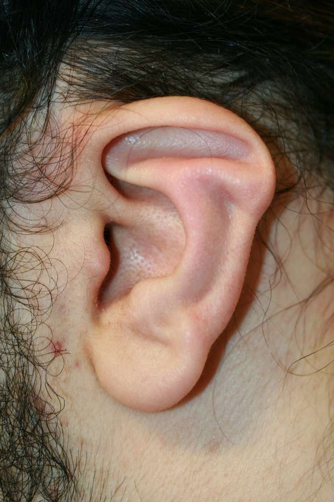 Stahl's bar
 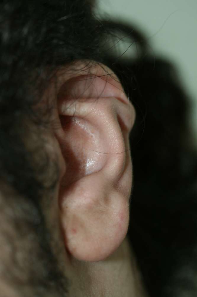 Stah's ear, oblique view
 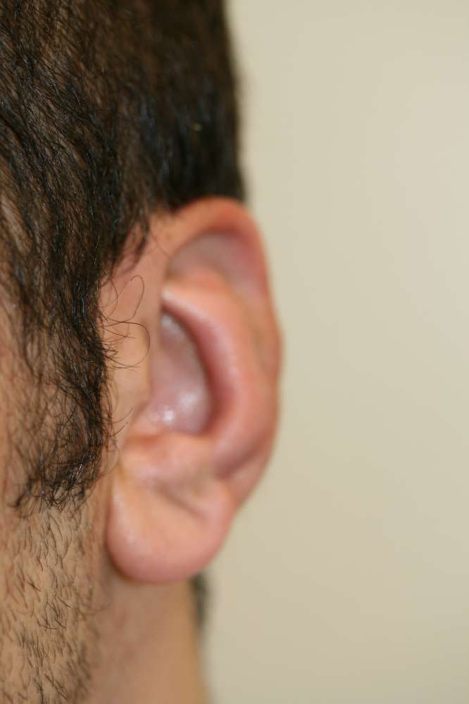 Stahl's bar correction
 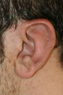 Stahl's ear correction - 6 weeks post surgery
 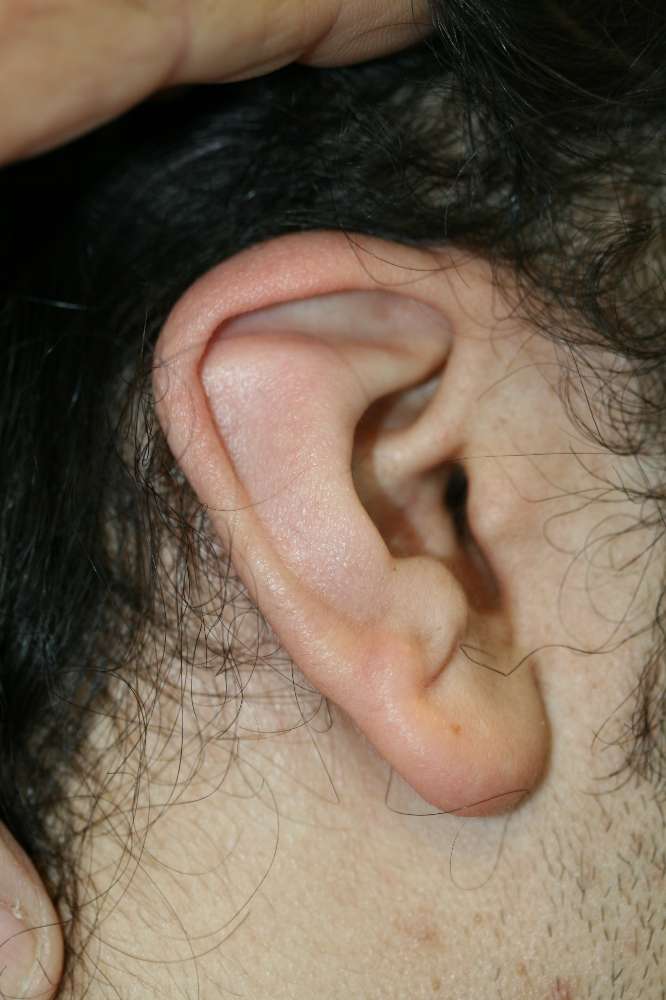 Stahl's ear, triangular ear shape
 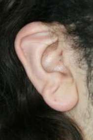 Oblique view of Stahl's ear, before surgery, showing small lop element
 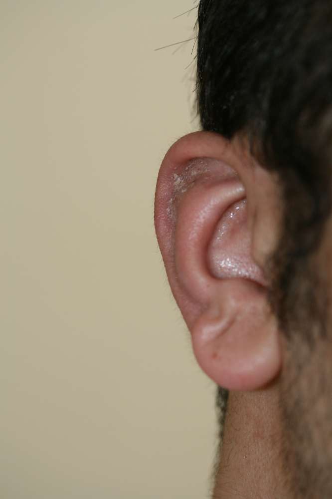 Stahl's bar correction - oblique view
 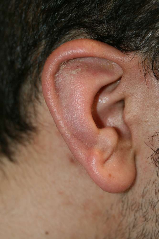 Stahl's ear correction - 6 weeks post surgery
 Pre-auricular skin tags
Pre-auricular skin tags are very common. Sometimes the tags are of skin only, but usually the tag contains a long tail of cartilage extending into the cheek.
If the "stalk" of the tag is relatively narrow, and the cartilage component relatively minor, tags can be removed soon after birth using an elegant clip device called a Liga Clip, which requires no anaesthetic. The clip cuts off the blood supply to the tag, and after a few days, the tag shrivels and separate. The sooner after birth the clip is applied, the better, and parents considering the removal of their new baby's pre-auricular deformity are recommended to make an early appointment.
Tags with a substantial cartilage core are best treated by excision of the skin tag and cartilage spindle under general anaesthetic, preferably when baby is over the age of 2 or 3 years.

Cryptotia
Cryptotia (crypt = hidden, otia = ear) is a hidden ear. The condition is obvious at birth and can affect one of both sides.
Sometimes only the lower two-thirds of an ear is visible and the groove above the ear seems lost. In fact, the top part of the ear, (the upper pole) is tethered to the side of the head, and hidden under the skin.When the ear is gently pulled away from the side of the head, the top of the ear can be seen, but it is usually reluctant to stay in a normal position.
The problem can be corrected soon after birth using EarBuddies™ Splints; if the opportunity is missed, then surgery is possible, preferably after the age of at least five years.
Ear Clefts
Clefts of the ear and ear lobe occur when two or more of the hillocks which form the ear during a baby's development in the womb fail to join together normally. Patients might complain that the ear looks like it has a "gap" in it, or that it is " missing a chunk", for example, where the earlobe meets the side of the head. The missing tissues can be restored using grafts or flaps to give a normal-looking ear.
Ear POsition problems - ear too low or too upright
Problems of ear position can also be remedied by surgery. The most attractive ears begin about one ear length behind the outer corner of the eye, and slope backwards in the same line as the nose. A change in this “angle” of the ear can confer unusual character - a very vertical ear appears abnormally formal, whereas one which slopes back too much can appear “drunk” or wayward. An ear which is too low or too close to the face can make its owner appear less intelligent. An injudicious face-lift can cause the ear to be pulled forwards and downwards, sometimes almost onto the cheek. This means that the scarring which can result is also brought into full view. Sometimes the ear lobes only are affected, either being pulled forwards, or made to stick out after a face-lift. All of these problems are correctable, but best avoided in the first place.
A procedure called auropexy can change the position of the ear in the same way as a mastopexy corrects drooping breasts. The misplaced ear is literally lifted back and re-sited in the correct position. For post face-lift patients, this has the additional benefit of improving the original lift.
PITS AND SINUSES
Pre-auricular sinuses can be difficult to excise and may recur after treatment. Very occasionally the sinuses track deeply near to the facial nerve. They often occur on both sides and frequently cause no trouble. However, if recurrent infections occur which fail to settle with antibiotics, surgical excision is appropriate.
Conchal crus
The conchal bowl is the cupped part of the ear that leads to the "ear hole". Sometimes the bowl is distorted by a ridge called a conchal crus, which often blocks the ear hole (auditory meatus), prevents the easy wearing of in-ear devices and spoils the appearance of the ear,
A conchal crus can be corrected in babies using Early Ear Correction with EarBuddies. In later life, the bar can be removed surgically.
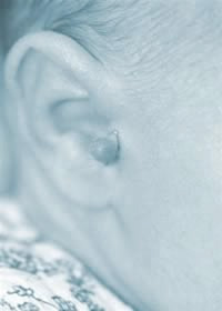
Darwin's Tubercle
A bump on the helical rim, on either one or both sides,is a common finding, often referred to as a Darwin’s tubercle. The lump is of normal ear cartilage, and is non-malignant. It has no health consequences, but is can give the ear an odd appearance, and can make the ear look as though it sticks out when it does not.
A Darwin's tubercle can be removed surgically, usually as an out-patient procedure under local anaesthetic.
 A nodular Darwin's tubercle
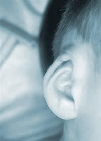
 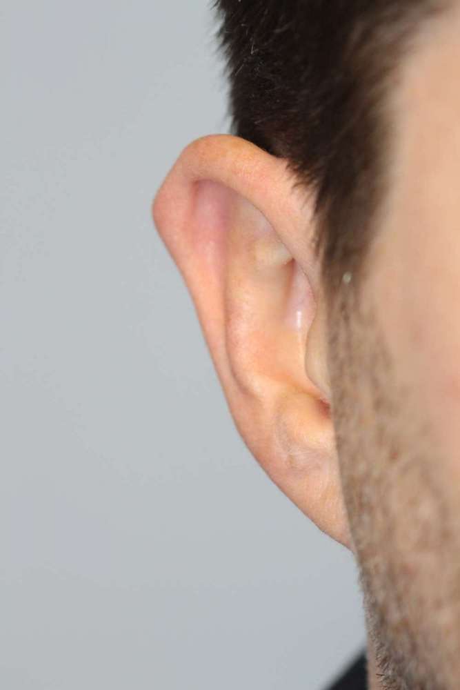 A more diffuse Darwin's tubercle
 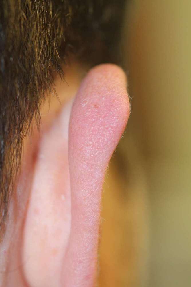 A more diffuse Darwin's tubercle from behind
 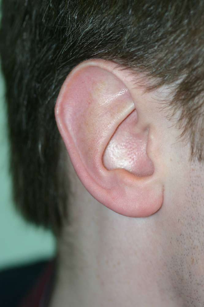 Large ear with small Darwin's tubercle
 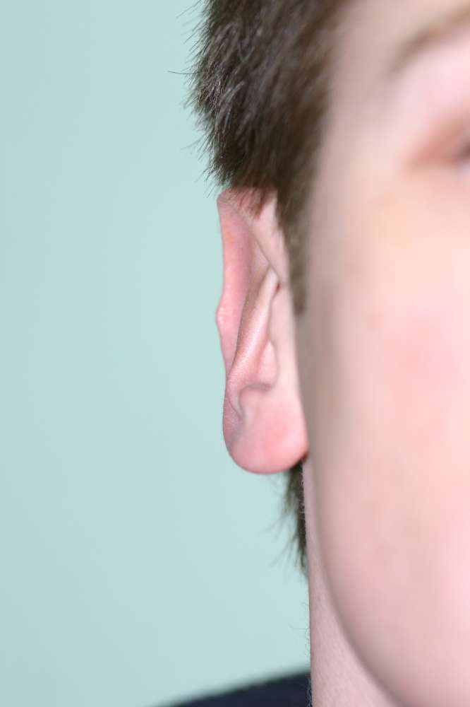 Darwins tubercle more obvious from the front
 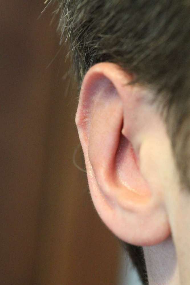 Darwin's tubercle in oblique view
 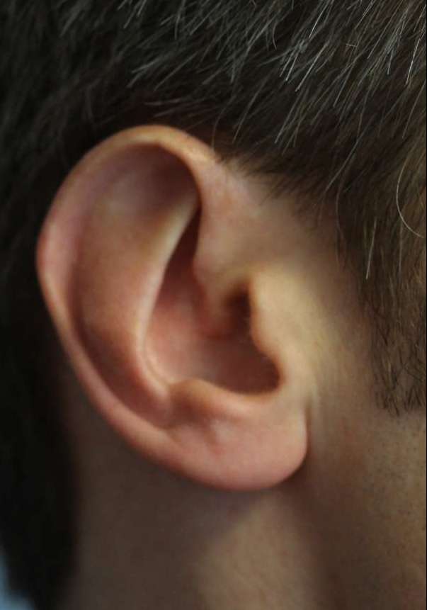 Smaller ear and Darwin's tubercle removed